
Can you really outrun cancer?
Meet the woman who is creating novel 3D models to try and find out


23 January 2020
When Dr Mhairi Morris’ nine-year-old daughter asked her across the breakfast table if it was possible to “catch cancer”, the School of Sport, Exercise and Health Sciences academic laughed and joked “of course you can’t, it’s far too fast”.
This witty one-liner got her thinking though, could it be possible to “outrun” the big C? How much of an impact does diet and lifestyle have on cancer risk and prevention?

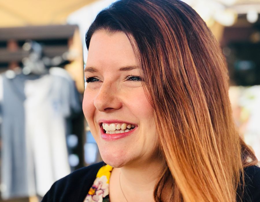
Dr Mhairi Morris, SFHEA - Lecturer in Biochemistry, School of Sport, Exercise and Health Sciences
Dr Mhairi Morris, SFHEA - Lecturer in Biochemistry, School of Sport, Exercise and Health Sciences
Mhairi is determined to find out and as part of her mission to do so, she is developing novel 3D models to try and understand the disease.
She discusses her research with VOLUME and her hopes for the future of cancer analysis.
WE ARE NOT MICE
“We are not mice!”, Mhairi tells a room of leading sport exercise experts as part of her talk at the National Centre for Sport and Exercise Medicine and Faculty of Sport and Exercise Medicine’s inaugural ‘Why is Moving Medicine?’ conference.
It may seem like an obvious statement, yet in the world of cancer research, mice are still used to grow tumours in a bid to mimic what is happening in human patients.
“Even if you put a human tumour in a mouse, it is surrounded by different types of mouse cell types – what we call ‘mouse stromal cells’ – therefore it’s not reflective of the human tumour microenvironment.
“We need to be looking elsewhere if we want to better understand this disease.”


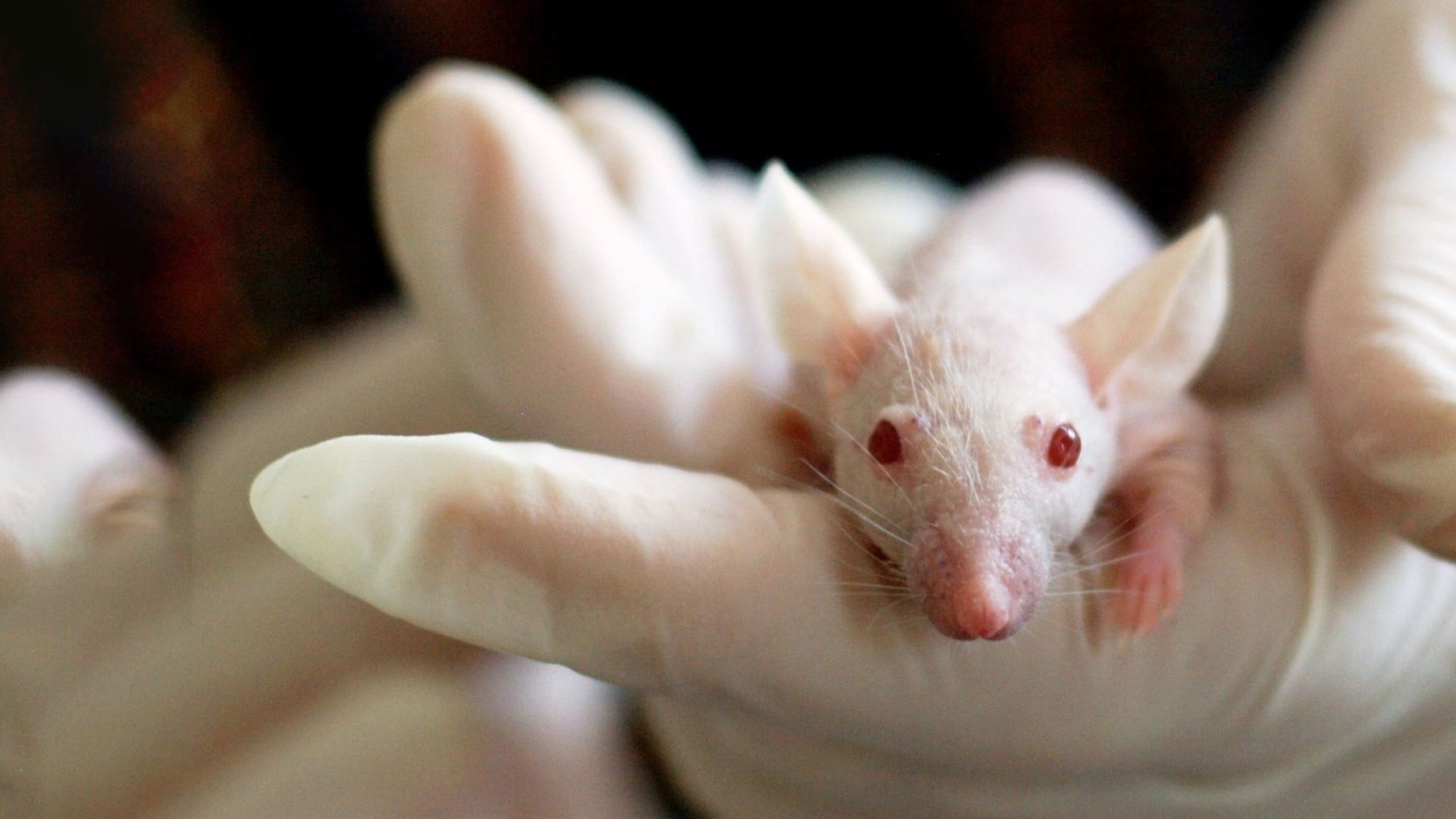
MOVING AWAY FROM MICE
Since graduating with a degree in Medical Biochemistry in 2004, Mhairi has been dedicated to researching and understanding different cancers.
Not a fan of the mouse models, she first started to explore what can be learnt from 2D monolayer cultures – cells that are grown in a dish, flat and side-by-side, under controlled conditions.
She had many successes with the models and revealed important new information about the viral oncoprotein latent membrane protein 1 (LMP1), which is thought to cause cancer in certain virus-associated cancers, including nasopharyngeal cancer (which affects the upper part of the throat behind the nose).
However, Mhairi found the models to be limited in what they can tell us about cancers.
She concluded that that they are not representative of what happens in the body and it is almost like 'playing football lying on the ground side-by-side – possible, but rubbish'.
In 2013, after returning to academia following maternity leave and a career break, Mhairi decided to turn her attention elsewhere.
She started to explore the wonderful world of 3D in vitro models.
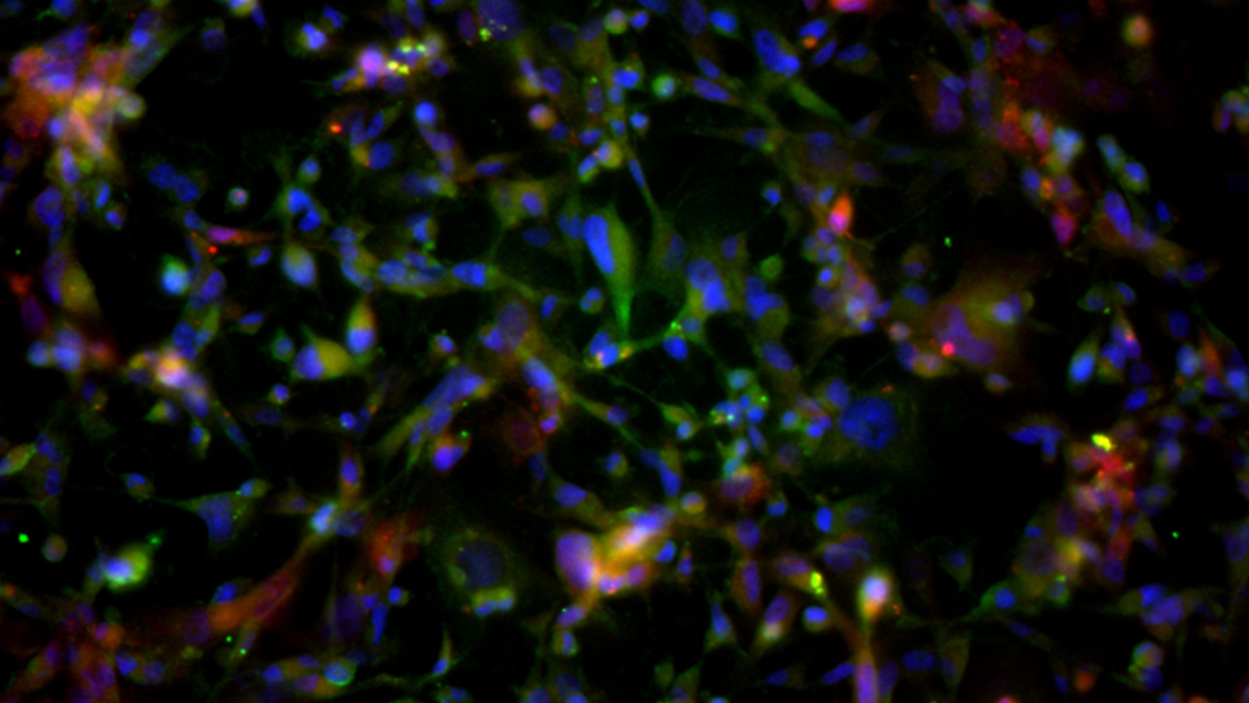
GROWING CELLS IN A 3D ENVIRONMENT
3D in vitro models are cell-based models of disease that are created in the lab (in vitro) and are used to investigate the inner workings of the cells that make up a tissue or organ.
They are like miniature tumours and can be built in a petri dish using all the different cell types that you’d find in an in vivo cancer (in the body).
They are used in a variety of health research areas – for example, experts have created 3D brain models to explore drug delivery, 3D organs have been printed, and bone structures have been recreated in vitro.
They are growing in popularity because they overcome the limitations of other methods and are therefore more representative of what happens in the body.
“Cells are in contact with each other on all surfaces in a 3D model,” Mhairi explained, “So you can mimic interactions, not only with other cells of the same type, but with other cell types, structures and tissues, and get a more accurate idea of how they'll behave in response to an intervention.
“For example, lots of clinical trials of drugs fail at the last hurdle because how a drug interacts in 2D cell studies and animal studies is just not the same as how it may interact with a real human.
“It’s also worth noting that the costs of developing transgenic mice and all the associated costs of animal testing is astronomical.
“By developing decent 3D models, we can potentially cut the massive costs involved in clinical trials, because in theory, the drug interactions with cells cultured in 3D will be more like what would actually happen in a human being.”
These models are a rapidly growing area in cancer research but not every stone has been turned over – especially when it comes to looking at the relationship between cancer and exercise.


A 3D CANCER MUSCLE MODEL THAT CAN REPLICATE THE EFFECTS OF EXERCISE
When Mhairi joined Loughborough University in 2017, she decided to shift her research focus to understanding how diet and lifestyle impact cancer risk and prevention.
Mhairi is developing brand new 3D models of cancers that have the potential to shed new light on cancer behaviour.
She is creating novel and, in some cases, never-been-done-before models that combine 3D tumours with other cell types, including 3D muscle tissues.
Mhairi oversees and works with two PhD students – one of whom, Mj Brown, is dedicated to researching and perfecting a 3D model of breast cancer and mesenchymal stem cells (or MSCs - a type of stromal cell that cancers can recruit to ‘do their bidding’) and has recently published a paper on the topic.
The next stage of the research will look at the impact of exercise on this cancer-stromal cell model, and how it affects the behaviour of MSC cells as they interact with breast cancer cells in vitro.
When asked why the link between exercise and cancer is an important research area, Mhairi said: “With cancer incidences on the rise, and almost half of all cancers being preventable through diet and lifestyle changes, it's important that we understand exactly how these interventions work to reduce cancer risk.”
She continued: “The difficulty with exercise science is that human beings exist in a finely tuned balance, and when one system is altered (e.g. when we exercise), everything else changes too in order to maintain that balance (called ‘homeostasis’), so it's really hard to define exactly what is having the effect on cancer growth and spread when a group of individuals are asked to take part in an exercise programme.
“In addition to this, we are all unique and will naturally respond differently to a given exercise intervention, so this adds another layer of complexity.
“If we had a system that mimics what goes on inside the body, but eliminates all these other variable factors (like the effect of other physiological systems and individual variation), then we can begin to unpick the tangled web of interconnected factors that may have an impact on tumour growth and spread.”
“It might even be that exercise improves the effectiveness of certain drugs that cancer classically develops a resistance to, and that could be really exciting!"


LOOKING TO THE FUTURE
Mhairi hopes the findings from the 3D models will one day be used to benefit real cancer patients.
“Ultimately, we want to take the results from our in vitro studies into cancer patients, safely, once we've identified the exercise-induced factors that may play a role in preventing primary tumours from spreading and becoming secondary tumours, which are the biggest killers in cancer”, she said.
“It might even be that exercise improves the effectiveness of certain drugs that cancer classically develops a resistance to, and that could be really exciting!
“We have had some promising findings in different areas but we've a long way to go before we can get excited – watch this space!”